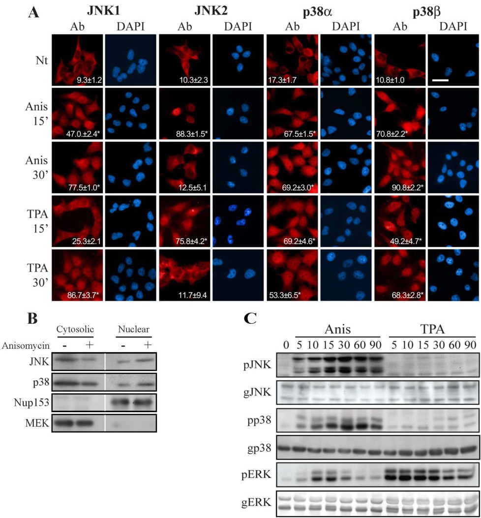
Fig. 1. JNK1/2 and p38α/β translocate into the nucleus independently of their activatory phosphorylation. (A) Fluorescent microscopy demonstrate the stimulated nuclear translocation of JNK1/2 and p38α/β. HeLa cells were grown on slides to 70% confluence, serum starved (0.1% FBS, 16 hr), and then stimulated with anisomycin (Anis, 0.5 µg/ml) or TPA (250 nM) for the indicated times, or left untreated. Cells were fixed, and stained with anti JNK1, JNK2, p38α or p38β Abs as indicated, and DAPI to detect nuclei. Percent cells with >40% nuclear staining and standard error was determenined by counting six distinct fields of 20 cells each from two distinct experiments. * - P< 0.05 compaired to non-stimulated cells. The bar in the upper right panel is of 20 µM. (B) Subcellular fractionation confirms the nuclear translocation of JNK1/2 and p38 α/β. Serum-starved HeLa cells were either stimulated (Anis, 0.5 µg/ml, 15 min) or left untreated, and then harvested. Subcellular fractions of cytosolic and nuclear were produced as described under materials and methods, and the fractions were subjected to Western blot analysis with the indicated Abs. (C) The nuclear translocation of endogenous JNK1/2 and p38α/β is independent of their phosphorylation. HeLa cells were grown to 70% confluence, serum starved, and then stimulated for the indicated time points. Cell extracts were produced and subjected to Western blot analysis with the indicated Abs. The blots were developed with NBT/BCIP or ECL. The experiments were reproduced with very similar results three times.